
Anti-GFAP
Rabbit Polyclonal Antibody
Catalog No. G28-563R
| Catalog No. | Pack Size | Price (USD) | |
|---|---|---|---|
| G28-563R-100 | 100 ug | $695 | |
| G28-563R-BULK | BULK | Contact Us |
Rabbit Polyclonal Antibody
Catalog No. G28-563R
| Catalog No. | Pack Size | Price (USD) | |
|---|---|---|---|
| G28-563R-100 | 100 ug | $695 | |
| G28-563R-BULK | BULK | Contact Us |
Overview:
Glial Fibrillary Acidic Protein (GFAP) was discovered as a major fibrous protein of multiple sclerosis plaques (1). It was subsequently found to be a member of the 10nm or intermediate filament (IF) family, specifically the IF family Class III, which also includes peripherin, desmin and vimentin. GFAP is strongly and specifically expressed in astrocytes and certain other astroglia in the CNS, in satellite cells, peripheral ganglia, and in non-myelinating Schwann cells in peripheral nerves. In many damage and disease states GFAP expression is heavily upregulated in astrocytes. In addition, neural stem cells frequently strongly express GFAP. Point mutations in the protein coding region of the GFAP gene lead to Alexander disease which is characterized by the presence of abnormal astrocytes containing GFAP protein aggregates known as Rosenthal fibers (2).
References:
1. Bignami A, Eng LF, Dahl D, Uyeda CT. Localization of the glial fibrillary acidic protein in astrocytes by immunofluorescence. Brain Res. 43:429-35 (1972).
2. Brenner M, Johnson AB, Boespflug-Tanguy O, Rodriguez D, Goldman JE and Messing A. Mutations in GFAP, encoding glial fibrillary acidic protein, are associated with Alexander disease. Nat Genet 27:117-20 (2001).
Specificity:
Recognizes the glial fibrillary acidic protein (GFAP)
Cross Reactivity:
Human, Mouse and Rat
Host / Isotype / Clone#:
Rabbit, IgG
Immunogen:
Recombinant and purified bovine GFAP.
Purification:
Affinity chromatography
Stability:
Store at 4oC (add 0.1% NaN3) for several months, and at -20oC for longer periods. For optimal storage, aliquot target into smaller quantities after centrifugation and store at recommended temperature. For most favorable performance, avoid repeated handling and multiple freeze/thaw cycles.
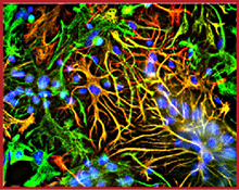
Sample Data:
Immunofluorescence showing specific labeling of GFAP (red) and vimentin (green) in cultured neurons and glia. Cells containing GFAP and vimentin appear yellow.
|
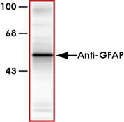
Sample Data:
Western blot of rat cortex lysate showing specific immunolabeling of the ~50k GFAP protein.
|
There are no related publications available for this product.
Neurobiology
STAY CONNECTED
Fax: 1-604-232-4601


 Toll Free: 1-866-954-6273
 info@signalchem.com